Abnormal hCG rise in an early pregnancy
Abnormal hCG rise predicts an unhealthy pregnancy; it is either a miscarriage or an ectopic pregnancy
The trend of hCG rise is the best predictor of the health of a pregnancy.
A healthy pregnancy at 6 2/7 weeks gestation is expected to have a heart rate of > 100/min.
Ms. McKesson was told that she had a miscarriage after having an ultrasound today – the fetal heart activity, observed one week ago, is no longer present. She accepted the diagnosis with composure - she was given a heads-up more than a week ago.
After the first positive hCG, I normally monitor pregnancies closely: 1) serum hCG twice weekly (Monday and Thursday or Tuesday and Friday) for two weeks, 2) a transvaginal ultrasound 2-3 days after the last hCG.
Based on the above, at the time of the first hCG test, the gestation age of pregnancies achieved via IVF, IUI, or timed intercourse treatment is approximately 4 weeks. By the time of the first ultrasound, the pregnancy is 6 2/7 weeks or more, when fetal cardiac activity is expected. The gestation age of pregnancies from spontaneous conception is more difficult to determine (due to unreliable last menstrual period). Despite this, I still order four hCG tests (over two weeks span) before ordering the ultrasound. Fewer blood is drawn, and an ultrasound is ordered when the hCG level reaches 1,500 mIU/mL.
The benefits of close monitoring include 1) giving patients a realistic expectation of the pregnancy, and 2) identifying abnormal pregnancies such as miscarriage or possible tubal pregnancy. The latter offers the benefit of medical treatment for presumed ectopic pregnancy.
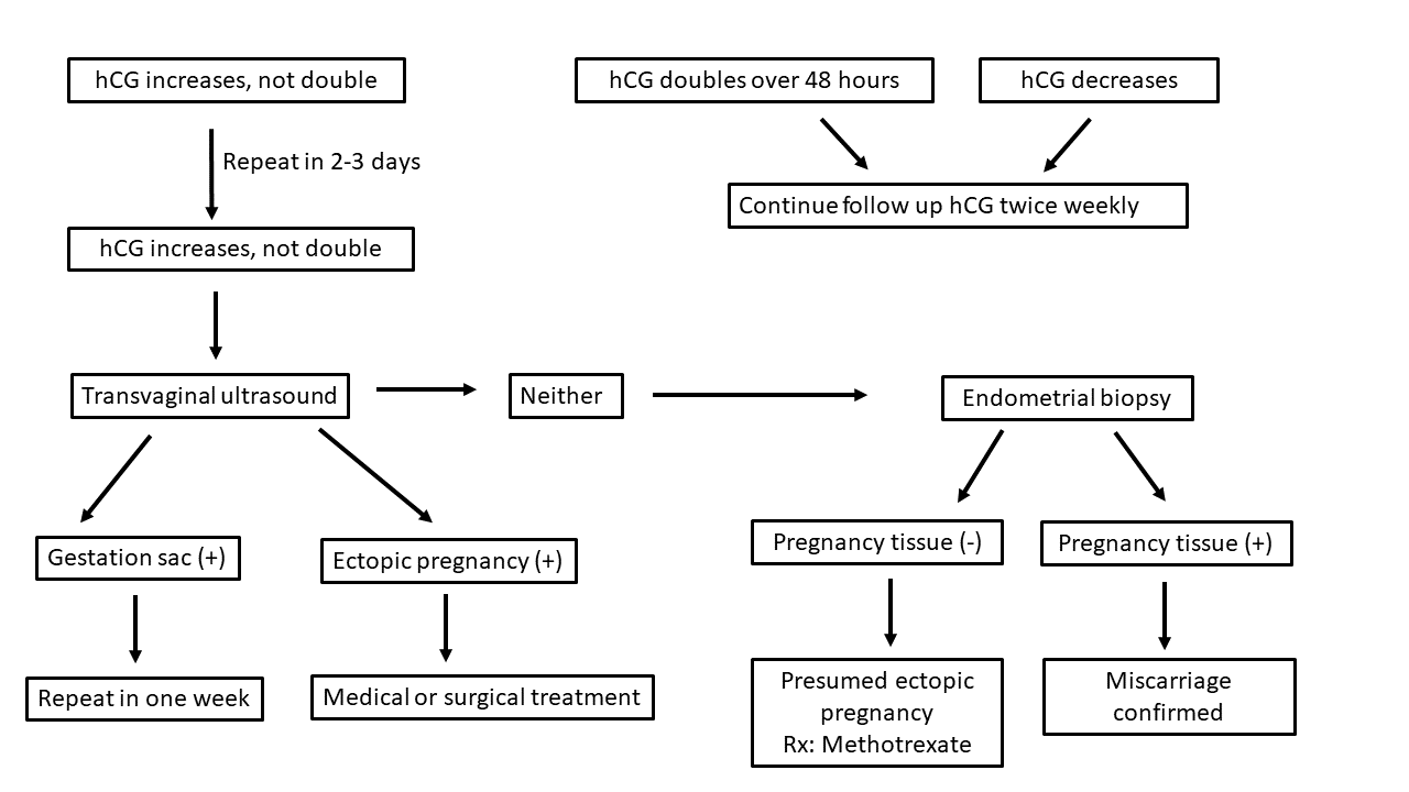
To avoid inter-laboratory variation and to have ease of comparison, I encourage all blood tests by the same laboratory. There are three scenarios (diagram below): #1, hCG rises more than two-folds (table below, left), #2 hCG decreases, and #3 hCG rises, but does not double. Ms. McKesson, who conceived by insemination, has the third scenario. Miscarriage poses no health concern, but ectopic pregnancy has grave consequences.
In the case of the third scenario (diagram below), I normally observe hCG falls off the curve twice (thereby, ruling out laboratory error) before ordering a transvaginal ultrasound. I am looking for a gestation sac or anything in the adnexa suggestive of ectopic pregnancy. If the ultrasound shows neither, I will offer the patient an endometrial biopsy and request a next-day result. The patient will be on call the next day for methotrexate injection (for presumed ectopic pregnancy) - if the pathologist does not see evidence of pregnancy in the sample
An abnormal hCG rise was brought to Ms. McKesson’s attention on 8/23 when the rise of hCG fell off the curve for the first time (McKesson’s values are on the left of the table below; values of a healthy pregnancy are on the right). She was told she may have a miscarriage or an ectopic pregnancy; she was given ectopic precautions. By 8/25, the hCG rise fell off the curve for the second time. A transvaginal ultrasound was scheduled for 8/29 with contingent endometrial biopsy.

The 8/29 ultrasound showed “single viable intrauterine pregnancy @ 6w1d-size is slightly small for dates. FHR-95bpm. The gestational sac measures 8.5mm consistent with 5w5d-slightly small for dates. The yolk sac measures 4.2mm-wnl. No evidence of a subchorionic hemorrhage. CL cyst on the RT ovary (8/29)” But by 9/6, the heart had already stopped. Ms. McKesson miscarried soon after; her hCG decreased to < 5 mIU/mL.
Ms. McKesson received the diagnosis of miscarriage well prepared because she was warned of the possibility when her serial hCG fell off the curve. She was given another warning when the ultrasound showed a heart rate of 95/min. She was warned twice that the pregnancy may not end well; she was prepared.
Two observations from this case are worth emphasizing: 1) hCG rise predicts best the health of an early pregnancy, 2) a < 100/min heart rate at 6 2/7 weeks gestation is an ominous sign.
It is worth mentioning that the “normal” fetal heart rate varies with the gestation age. Fetal cardiac activity can be observed by a transvaginal ultrasound at 5 5/7 weeks gestation; the rate is usually lower than 100/min. But by 6 2/7 weeks gestation, a healthy pregnancy’s heart rate should be > 100/min.


